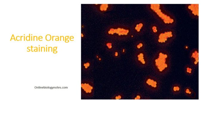
Principle:
Acridine orange is a fluorochromatic dye which binds to nucleic acids of bacteria and other cells that causes deoxyribonucleic acid (DNA) to fluoresce green and ribonucleic acid (RNA) or single stranded DNA to fluoresce orange-red under UV light. It was first described by Strugge and Hilbrich in 1942. When buffered at pH 3.5 to 4.0, acridine orange differentially stains microorganisms from cellular materials. It has been recommended for the rapid identification of Trichomonas vaginalis, yeast cells, and clue cells in vaginal smears. Intracellular gonococci, meningococci, and other bacteria particularly in blood cultures are also detected. In clinical samples, Bacteria and fungi stain bright orange whereas, Human epithelial and inflammatory cells and background debris stain pale green to yellow. Nuclei of activated leukocytes stain yellow, orange, or red due to increased RNA production resulting from activation. Erythrocytes either do not stain or appear pale green.
The acridine-orange stain is an optional stain that can be helpful in detecting organisms not visualized by Gram stain, often due to a large amount of host cellular debris.
Requirements:
- Acridine orange acid stain (Store at 15 to 30 °C in the dark).
- Alcohol saline solution(Absolute Methanol)
- Microscopic glass slides
- Heat block at 45 to 60° C (optional)
- Fluorescent microscope
- Immersion oil
Quality control:
- Examine the acridine orange staining solution for color and clarity. The solution should be clear, orange, and without evidence of precipitate.
- Each time of use, stain a prepared slide of known bacteria, such as Escherichia coli mixed with staphylococci, and examine for the desired results. Record results and refer out-of-control results to the supervisor.
- Result: Gram-negative rods and gram-positive cocci are fluorescent (orange) and Background is non-fluorescent (green-yellow).
Procedure for Acridine orange staining:
- Prepare a smear of the specimen on a clean grease free glass slide
- Spread the specimen thinly with a sterile stick.
- Allow to air dry, or speed dry by placing the smear on the heating block.
- Alcohol fixing: Fix the smear with methanol by flooding the slide, draining the excess; allow to air dry.
- Flood slide with acridine orange stain for 2 min.
- Drain the excess stain and rinse thoroughly with tap water.
- Allow to air dry.
- The slide may be gently blotted on a clean sheet of filter paper or paper towel to decrease drying time.
- Observe the smear on the fluorescent microscope. Look for the distinct morphology of bacteria or fungi. No coverslip is needed
Result interpretation:
- Bacteria and fungi appears bright orange.
- Trichomonas vaginalis trichomonads appears Orange or red with yellow-green nucleus
- The background appears black to yellow green.
- Human epithelial and inflammatory cells and tissue debris appears pale green to yellow.
- Activated leukocytes will appears yellow, orange, or red, depending on the level of activation and amount of RNA produced, whereas erythrocytes either do not fluoresce or fluoresce pale green.
- Ureaplasma and Mycoplasma can be visualized on acridine staining.
Limitations of Acridine orange stain:
- Acridine orange also stains nucleus or granules of activated leukocytes, and certain types of other cell debris, therefore, it is difficult to separate them from microorganism. However, these may be differentiated from microorganisms on the basis of morphology.
- Acridine orange staining does not distinguish between gram-negative and gram positive organisms.
- Intracellular organisms may be more difficult to see by the acridine orange stain, due to the staining of cellular nuclei.
- The acridine orange stain will be used to stain some Gram negative which are poorly visualized by Gram staining.
- The sensitivity of the acridine orange smear is approximately 104 bacteria/ml
