Antibody mediated immune response
- The antibody mediated immune response is also known as humoral immunity.
- It is the host defense mechanism that are mediated by antibody present in plasma, lymph and tissue fluids.
- It is the type of adaptive immune system responsible for defense against extracellular microbes, microbial toxins and foreign macromolecules.
- The cells involved in AMI are B-lymphocytes, TH cells and phagocytic cells.
Mechanism of Antibody mediated immune response:
- The effector response of antibody mediated immune response completes in three steps:
- Activation and proliferation of B-cells to produce antibody
- Class switching of antibody
- Antibody mediated removal of antigen
Step I: Activation and proliferation of B-cells
- When the B-cell development and maturation completes in bone marrow, they are exported to the peripheral lymphoid tissues.
- When the mature B-cell in periphery is first exposed to antigen, they are activated by extensive cross-linkage of antigens to the mIg receptor on B-cells.
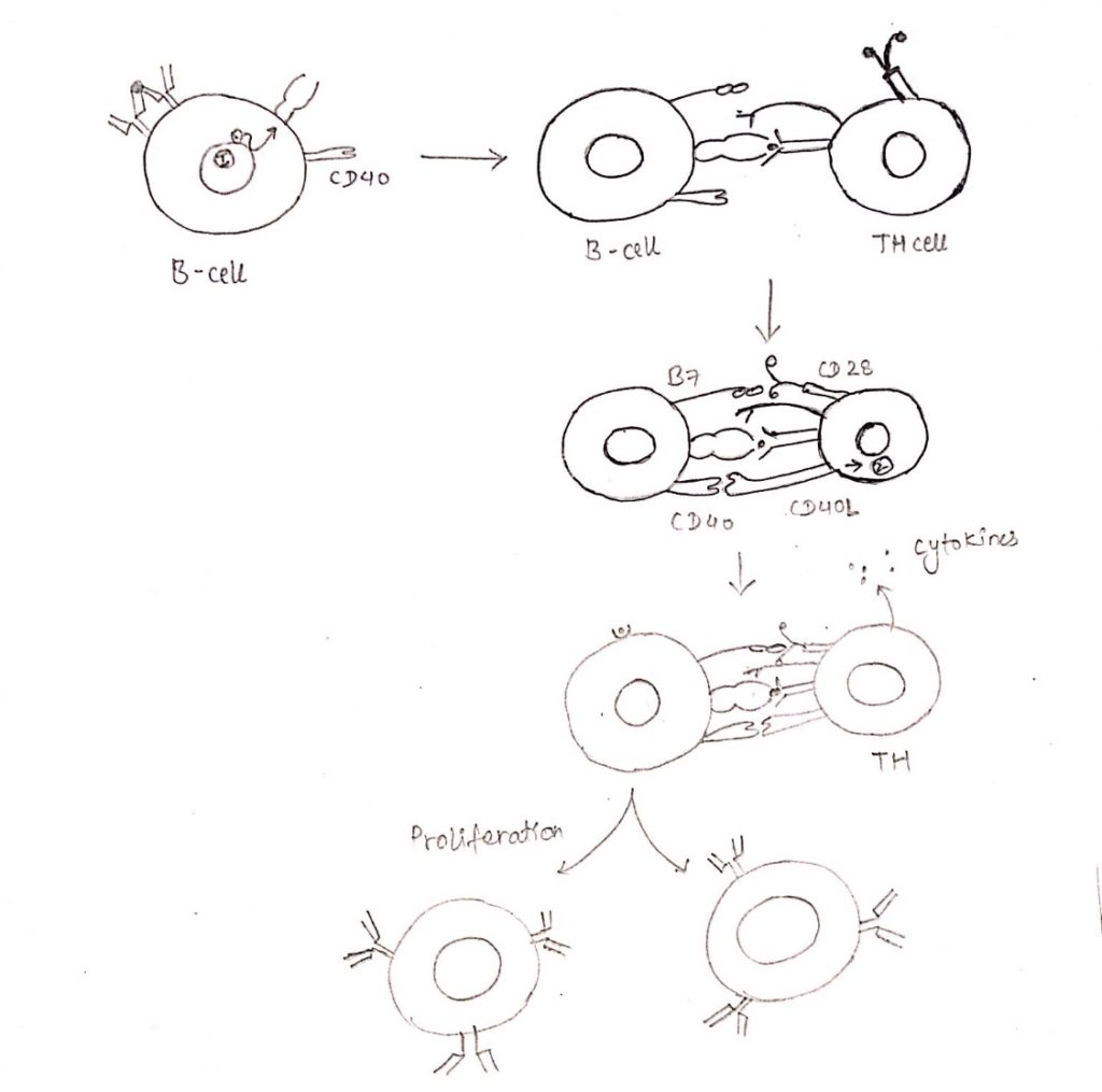
- The cross-linking of Ag with mIg generates signal 1 which leads to internalization of Ag-Ab complex by endocytosis and increase expression of class II MHC molecule with antigenic peptides and co-stimulatory receptor B7.
- Then the antigen presented with MHC-II on B-cell membrane is recognized by TCR of TH cell.
- This interaction plus co-stimulatory signal generated by B7-CD25 interaction activates the TH cells.
- The activated TH cell then begins to express CD40L.
- The interaction of CD40 and CD40L provides signal 2.
- The signal 2 along with co-stimulatory signal provided by B7-CD28 interaction stimulates the B-cell to begin expression of receptor for various cytokines.
- The binding of cytokines released by the TH cell to cytokines receptors in B-cell sends signals that supports the progression of B-cell to DNA synthesis and for the differentiation of B-cell into antibody producing plasma cell and memory cell.

Step II: Class switching of antibody
- In response to CD40 engagement and cytokines, some of the progeny of activated IgM and IgD expressing B-cell undergo the process of heavy chain isotype switching.
- It leads to the production of antibodies with heavy chain of different classes such as Ƴ, α and ε.
- Isotype switching occurs in peripheral lymphoid tissues.
- CD40 signaling and cytokinin produced by TH cell play essential role in regulatory switching to particular heavy chain isotypes.
- The major mechanism by which CD40 signals induce isotype switching is the induction of AID (Activation induced Deaminase) gene downstream of CD40.
Step III: Antibody mediated removal of antigen
- Antibody mediated removal of antigens consists of a number of effector mechanism.
- It needs the participation of various cellular and humoral component of immune system involving phagocytes and complements.
- Neutralization
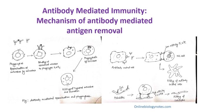
- Antibody mediated opsonization and phagocytosis
- Antibody dependent cell mediated cytotoxicity (ADCC)
- Complement system
i. Neutralization of microbes and microbial toxins:
- The first step for removal of Ag is neutralization of microbes and toxins.
- Neutralization is the process by which antibodies against microbes and microbial toxins blocks the binding of these microbes and toxins to cellular receptor, thus inhibiting the infections.
- Neutralization of microbes by antibodies may occur by different ways.
- Microbes enter the host cells by binding of their particular surface molecule to host cellular receptor antibodies then bind to these microbial structures and interfere with the ability of microbes to interact with cellular receptors. This interaction is called steric hindrance.
- Very few antibodies bind to microbes and induce conformational change in the surface molecules that prevent the microbes from interacting with cellular receptor. This interaction is called allosteric effect of antibodies.
- Microbial toxins also cause pathogenic infection by binding to specific cellular receptors. Antitoxin (antibodies) sterically hinder the interaction of toxin to cellular receptor of host cell and prevent the tissue from injury and disease.
ii. Antibody dependent cell mediated cytotoxicity:
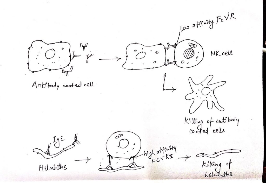
- NK cells and other leukocytes bind to antibody coated cells by the receptor and destroy these cells.
- This process is termed as antibody dependent cell mediated cytotoxicity.
- FcƳR III is present on the NK cells which binds to antibody coated cells.
- Since, FcƳR III is a low affinity receptor, it can bind only IgG bound infected cells but does not bind circulating monomeric IgG.
- Thus, ADCC takes place only when the target cell is coated with antibody molecules.
- Interaction of FcƳR of NK cell with IgG coated target cells, activates the NK cell to synthesize and secrete cytokines such as IFN-Ƴ and discharge the contents of their granules.
- This leads to lysis of antibody coated cells.
- Eosinophils, mediate a special type of ADCC directed against helminthic parasites.
- Helminths are too large to be engulfed by the phagocytes and their integument is relatively resistant to the microbicidal products of neutrophils and macrophages.
- They can be killed by a basic protein present in the granules of eosinophils.
- IgE coats the helminths and eosinophils can then bind to IgE through FcƳRI.
- The binding activates eosinophil to release their granules content which results in the killing of the helminths.

iii. Antibody mediated opsonization and phagocytosis:
- Mononuclear phagocytosis and neutrophils express a variety of surface receptors that directly bind microbes and ingest them and cause intracellular killing and degradation.
- Mononuclear phagocytosis and neutrophils express receptors for the Fc portion of IgG antibodies that specifically bind antibody coated (opsonized) particles.
- A product of complement activation called C3b can also opsonize microbes and are phagocytosed by binding to a leukocyte receptor for C3b.
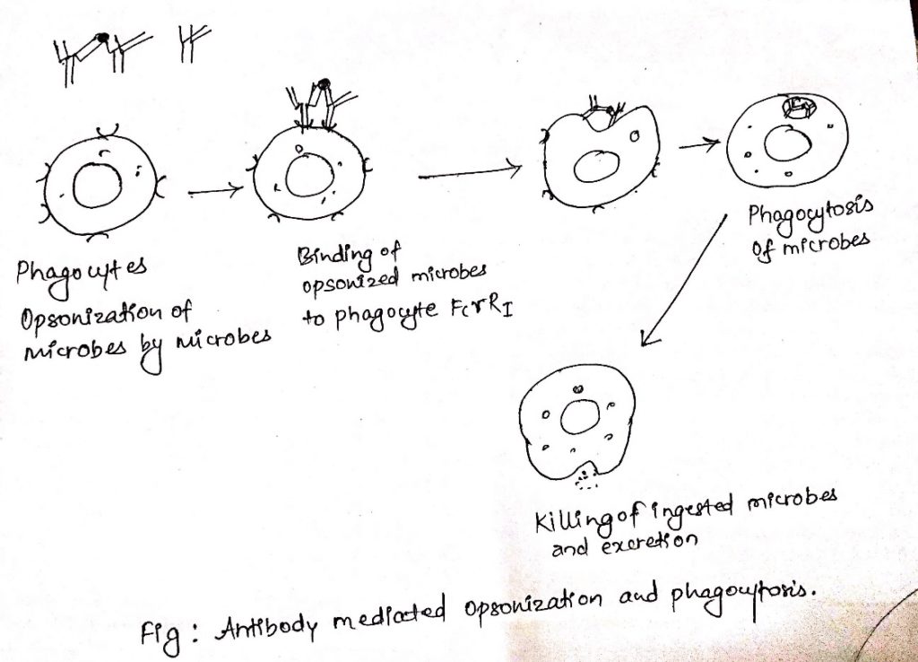
- The process of coating particles for phagocytosis is called opsonization.
- Antibodies and complements proteins that perform their function are called opsonins.
- Phagocytosis of IgG coated particles is mediated by binding of the Fc portion of opsonizing antibodies to FcƳR on phagocytosis.
- Antibodies bound to antigens from multivalent arrays and are bound by phagocyte FcR with much higher avidity than free antibodies.
- Binding of multiple FcR to antibody coated particle lead to engulfment of the particles and their internalization in phagocytic vesicles.
- These phagosomes fused with lysosomes and the phagocytosed particles are destroyed in phagolysosome.
- Phagocytosis is also activated by the signal transducted by binding of opsonized particles to FcƳR I.
- This signal result in activation of tyrosine kinase in the phagocytes, which stimulates production of various microbicidal molecules.
- Phagocyte activation leads to the production of enzyme phagocyte oxidase, which catalyse the intracellular generation of reactive oxygen intermediates that are cytotoxic to phagocytosed microbes.
- Too large microbes that cannot be phagocytosed are killed by the activation of leukocytes that is capable of extracellular killing by secretion of hydrolytic enzymes and reactive oxygen intermediates.

iv. Activation of Complement system:
- Complement system is a system of lytic enzyme found in blood.
- These lytic enzymes are usually inactive, they are activated only under particular condition to generate products that mediate various effector functions of complement.
- Activation of complement involves the sequential proteolysis of protein to generate newly assembled enzymes complex with proteolytic activity.
- Zymogens are termed as the proteins that gain proteolytic enzymatic activity by the action of other proteases.
- The process of sequential zymogen activation is an enzymatic cascade.
- Proteolytic cascade allows tremendous amplification because each enzyme molecule activated at one step can generate multiple activated enzyme at next steps.
- The products of complement activation become covalently attached to microbial cells surface or to antibodies bound to microbes and to other antigens.
