Structure and Functions of Human Digestive glands:
- The glands that secrete digestive juices for the digestion of food are termed digestive glands.
- Besides the number of gastric glands present in the lining of the stomach, there are many other related digestive glands that pour their secretions into the alimentary canal.
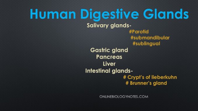
- The digestive glands include salivary glands, gastric glands, liver, pancreas, and intestinal glands.
1. Salivary glands:
- Three pairs of salivary glands are present.
- They are the parotid, submandibular and sublingual glands.
- Parotid glands:
- The largest of the salivary glands are parotid glands.
- They are located one on each side of the face, just below and in front of the ears.
- Parotid glands secrete saliva via a parotid duct into the mouth and help in mastication and swallowing,
- The secretion of each parotid gland passes through Stensen’s duct which opens into the mouth opposite the site of second upper molar tooth.
- Mumps is the disease caused by the viral infection of parotid gland.
- It is characterized by swelling, irritation, and pain.
- Submandibular glands:
- These are located embedded in the mucous membrane on the floor of the buccal cavity, under the tongue.
- Duct of these glands open into the sublingual part of the mouth under the tongue.
- It secretes mixture of the serous fluid and mucus.
- This secretion passes into the oral cavity via the submandibular duct or Wharton’s duct.
- Despite being much smaller than the parotid glands, it accounts for the production of 65-70% of saliva in the oral cavity.
- Sublingual glands:
- It is the smallest of all the salivary glands.
- It also includes the parotid and submandibular glands.
- It is located between the muscles of the oral cavity.
- It is different from other salivary glands as it lacks striated ducts, hence the saliva is secreted directly via the ducts of Rivinus.
- It is the only unencapsulated major salivary gland.
- Primarily, it secretes a thick mucinous fluid that lubricates the oral cavity and aids for swallowing, buffering the pH, initiate digestion, and dental hygiene.
Saliva and its fuctions:
- The salivary glands secrete saliva which is a viscous fluid.
- The saliva of man is a viscous, colorless, cloudy, and opalescent liquid.
- The optimum pH value is 6.8 with a range of 5.6 to 7.6 and specific gravity ranges from 1.002 to 1.008.
- It is uniformly secreted in small quantities to keep the buccal cavity moist.
- When food is present the rate of secretion is increased as the saliva helps both moistening the food and lubricating its subsequent passage through the alimentary canal.
- It also initiates digestion.
- The saliva contains 98.5 to 99% water and 1 – 1.5 % of a dense residue.
Enzymes of saliva:
- Saliva possesses a large number of enzymes such as amylase, lysosome acid phosphatase, aldolase, cholesterase, lysozyme, maltase, catalase, lipase, urease, and protease.
- Among them, amylase and lysozyme show physiological importance.
- Salivary amylase carries out the hydrolysis of starch and glycogen to maltose, isomaltose, dextrin and some glucose.
- Some particular complex polysaccharides present in the cell wall of different species of bacteria are hydrolysed by lysozyme, thereby killing and dissolving them.
Functions of saliva:
- The saliva is responsible for a number of functions:
- The dry food is moistened and facilitates swallowing by a lubricating action. It prevents desiccation of the oral mucosa, since water evaporates slowly from saliva.
- One of the enzymes present in saliva, salivary amylase, or ptyalin play a role in the digestion of starch.
- It keeps the mouth and teeth clean.
- The soluble substance such as sugar and salts get dissolved by it.
- It makes the food delicious to taste.
- The excretion of certain substances such as lead, mercury, and iodides take place in the saliva.
- It makes rapid articulation possible by facilitating movements of the tongue and lips.
- It enhances the sense of taste by acting as a solvent. The taste buds can be stimulated only when the substances having pleasant taste are actually present in the solution.
- It consists of three buffering systems, bicarbonate, phosphate, and mucin of which the bicarbonate is most important.
- When the salivary flow increases particularly at the time of eating, the concentration of bicarbonate and the buffering system rises.
2. Gastric glands:
- The wall of the stomach has various gastric glands.
- They are simple or branched tubular glands.
- About 2-3 litres of gastric juice is secreted daily by these gastric glands in adults.
- At least three different types of gastric glands are present in the gastric mucosa. These are: parietal cells (oxyntic cells), chief cells, and mucous cells.
- The parietal cells supply the hydrochloric acid of the gastric juice.
- The chief cells provide pepsin and other enzymes such as rennin and gastric lipase and the mucous cells secrete mucin.
- The Castle’s intrinsic gastric factor is secreted by the parietal cells that help in the absorption of vitamin B12.
- All these secretions altogether form an acidic gastric juice with pH=2 and low specific gravity ranging from 1.002 to 1.006.
- The gastric juice comprises about 0.5% of solid matter including sodium chloride with traces of potassium chloride and phosphates, mucin and enzymes pepsin, rennin and gastric lipase, hydrochloric acid and mucin.
- The secretion of gastric juice is under both nervous and hormonal control.
- Gastric secretion is triggered by the thought of food, the smell of food, chewing of food and contact of food with the stomach wall.
- The gastric glands also have two uncommon types of gland cells:
- Argentaffin cells that are normally situated at the base of the glands and secrete serotonin, a potent vasoconstrictor.
- Endocrine cells that are present in the pyloric antrum and produce gastrin. The gastrin triggers the secretion of enzymes and HCl.
3. Liver:
- It is the largest gland of the body.
- It generally weighs about 1.6 kg.
- It is chocolate colored and is located just behind the diaphragm on the right side of the upper abdominal cavity.
- The liver has two main lobes, the right lobe which is larger, and the left lobe along which is smaller with two small lobes, the quadrate lobe and the caudate lobe present behind the main lobes.
- Hepatic artery supplies to the liver.
- The blood is drained into the inferior venacava by the hepatic vein.
- On the undersurface of the right lobe of liver, a pear-shaped structure is located called as a gall bladder.
- The gall bladder serves as a reservoir that stores the bile juice secreted by the liver.
- The right and left hepatic ducts from the liver join to form a common bile duct or ductus choledochus.
- The common bile duct and pancreatic duct join to form the hepatopancreatic duct, near the duodenum.
- This opens in the duodenum via an opening guarded by the sphincter of Oddi.
- The dilation of short common hepatopancreatic duct forms hepatopancreatic ampulla or Vater’s ampulla.
- The ampulla opens into the duodenum.
- The bile duct is surrounded by a strong sphincter muscle of Boyden before it is joined by the pancreatic duct.
- When there is no food in the duodenum, this muscle closes.
- This forces the bile into the gall bladder through the cystic duct for storage.
Functions of the liver:
- The liver being a multipurpose organ, performs several important functions:
- Production of bile:
- Liver secretes bile which is an alkaline dark green-colored fluid having several organic and inorganic salts as well as some waste substances.
- The bile performs the following functions:
- It makes the chyme alkaline, better suited for the action of pancreatic juice.
- It is responsible for the emulsification of fats.
- It helps in removing the excretory products like bile pigments, inorganic salts, toxins, etc. from the body.
- It stimulates peristalsis.
- Some bile salts are necessary for the absorption of vitamin K and other vitamins soluble in fats.
- Bile acts as an antiseptic, therefore, it does not allow the growth and multiplication of bacteria.
- Regulation of blood sugar level:
- Liver regulates the blood sugar level (normal 90-120mg per ml of blood) either by the process of glycogenesis or by the glycogenolysis.
- Glycogenesis is the conversion of glycogen into glucose by the liver cells with the help of glucagon secreted by the pancreas.
- It is also a center for gluconeogenesis and glyconeogenesis.
- Lipogenesis:
- Liver also controls lipogenesis. The conversion of the excess of glucose and amino acids into the fats is termed as lipogenesis. It also takes place into the liver.
- Deamination:
- Liver decomposes the excess and harmful amino acids of blood into toxic NH3 and metabolically useful keto acids by the process of deamination in the presence of oxidase enzyme.
- In the liver cells, toxic NH3 is combined with CO2 to form less toxic urea by the process of detoxification.
- Liver also detoxifies the alcohol and converts it into acetaldehyde and then harmless acetyl CoA.
- Absorption and metabolization of bilirubin:
- Liver transforms hemoglobin of dead RBCs into bile pigments such as biliverdin and bilirubin which are egested out along with feces. Thus, liver helps in excretion.
- Aids in blood clotting:
- Liver produces an anticoagulant called heparin which prevents the coagulation in the blood vessels.
- Liver produces two main proteins fibrinogen and prothrombin which help in clotting of blood at injury to check excess of bleeding.
- Erythropoesis and hemolytic function:
- Liver acts as an erythropoietic organ. It forms RBCs in the foetus.
- Liver also acts as hemolytic organ. It breaks old RBCs.
- Immunological functions:
- Kupffer’s cells of liver act as phagocytes which feed on dead cells and bacteria by the phagocytosis process.
- Vitamin and mineral storage:
- Liver synthesizes vitamin A from beta carotene in the presence of an enzyme carotenase. Beta carotene is an orange yellow substance of carrot.
- Liver stores minerals like copper and iron, vitamins like A, D, E, K and B12, glycogen, fats and water.
- Maintains body temperature:
- Liver is the major heat producing centre of the body. Because of high metabolic activities of the liver, sufficient heat is generated, which is very important for maintaining the optimum body temperature.
- Blood reservoir:
- It serves as the second reservoir of blood.
- Angiotensinogen synthesis:
- Liver secretes a protein called angiotensinogen which helps kidney to maintain body fluid, osmoregulation.
- Blood filtration:
- It helps in eliminating several unwanted substances like carbolic acid, cresol, etc. from the blood coming from alimentary canal.
- It is the important seat of lymph formation.
- It also produces certain substances which check anemia.
4. Pancreas:
- The pancreas is an elongated and yellowish gland.
- It is located behind the stomach lying horizontally in the curvature of duodenum.
- It is about 12-15cm long and 2.5 cm wide and weighs about 60gms.
- It comprises of head, body and tail.
- The head is located in the curve of the duodenum, the body lies behind the stomach and the tail links the spleen.
- The smaller ducts within the pancreas form the main pancreatic duct.
- Into the hepatopancreatic ampulla (ampulla of Vater), the main pancreatic duct opens.
- In pancreas, an accessory pancreatic duct (duct of Santorini) is also present and opens directly into the duodenum.
- The pancreas is exocrine as well as endocrine in nature.
- A large number of branching tubules or lobules are present in the exocrine tissue called acini.
- Acini are embedded in connective tissue containing blood and lymph vessels, nerves and pancreatic ductules.
- The acinus comprises of cuboidal epithelial cells and secretes an alkaline pancreatic juice with pH 8.8.
- About 500-800 ml of pancreatic juice is secreted every day.
- The main pancreatic duct carries the pancreatic juice into the duodenum through the hepatopancreatic ampulla.
- The pancreatic juice is directly poured into the duodenum by the accessory pancreatic duct.
- The pancreatic juice contains sodium bicarbonate, three proenzymes: trypsinogen, chymotrypsinogen and procarboxypeptidase and several enzymes: pancreatic amylase, pancreatic lipase and nucleases such as DNAse and RNAse.
- The pancreatic juice aids in the digestion of starch, proteins, fats and nucleic acids.
- The activity of chyme caused by HCl is neutralized by the sodium bicarbonate.
- The endocrine tissue of pancreas consists of several group of cells, called Islets of Langerhans, found situated in between the acini.
- Each Islet of Langerhans comprises of the following types of cells.
- These secrete hormones to be poured in the circulating blood.
- i) Alpha cells (a-cells) are more numerous towards the periphery of islet and constitute about 25% of the Islet of Langerhans. These cells produce glucagon hormone which converts glycogen into glucose in the liver.
- ii) Beta cells (b-cells) are more numerous towards the middle of the islet and constitute about 60% of the islet and constitute about 60% of the islet of Langerhans. These cells supply insulin hormone that converts glucose into glycogen in the liver and muscles. Deficiency of insulin causes diabetes mellitus.
- iii) Delta cells (d-cells) are found towards the periphery of islet and constitute about 10% of islet of Langerhans. These cells produce somatostatin hormone which halts the secretion of glucagon by alpha cells and to some extent secretion of insulin by beta cells.
- iv) Pancreatic polypeptide cells (PP cells) are also found in the pancreas. Pancreatic polypeptide (PP) are secreted by these cells which inhibits the release of pancreatic juice.
- Thus, the pancreas serves two main functions:
- i) secretion of pancreatic juice which contains digestive enzymes and
- ii) production of hormones
5. Intestinal glands:
- They are numerous, microscopic glands present in the mucosa of small intestine.
- They are of two types i.e. crypts of Lieberkuhn and Brunner’s glands.
- Crypts of Lieberkuhn:
- These are simple tubular glands.
- They occur throughout the small intestine between villi.
- Digestive enzymes and mucus are secreted by it.
- These secretions are slightly alkaline with pH in the range of 7.5 to 8.0.
- They contain several types of cells that secrete mucus and a large number of enzymes.
- The secretion of these glands is termed as succus entericus or intestinal juice.
- Each day about 2-3 litres of intestinal juice is secreted.
- It is alkaline (pH 8.3) in nature and is poured into the intestine.
- The intestinal juice contains several enzymes: aminopeptidases, intestinal amylase, maltase, isomaltase, dipeptidases, limit dextrinase, sucrase, intestinal lipase, nucleotidases or nucleophosphatases, lactase, nucleosidases and enteropeptidase (enterokinase).
- These enzymes digest all types of food.
- Brunner’s glands:
- These are branched tubular glands.
- These are restricted to the submucosa of the duodenum.
- These glands secrete mucus.
- The mucus lubricates and sticks the food together.
- The digestive tract is therefore lubricated so that food quickly slips into it. This protects the duodenal mucosa from the stomach-derived acidic chyme.
- The mucus also protects mucosal cells from digestive enzymes.
- Along with the glands mentioned, the entire alimentary canal has mucous glands that produce mucus.
- The mucus lubricates the food and digestive tract and protects the mucosa of the stomach from damage.
