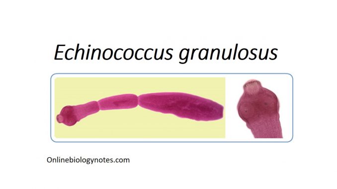
- Echinococcus granulosus also termed as the hydatid worm or Hyper tapeworm or Dog Tapeworm.
- It is a cyclophyllid cestode that parasites the small intestine of canids as an adult but which has vital intermediate hosts as livestock and humans where it results cystic echinococcus, also termed as hydatid disease.
Habitat:
- The adult tape worms are located affixed to the wall of intestinal mucosa of dogs and wild canines.
- The larval stage (hydatid cyst) is present in humans and other herbivorous animals.
Morphology of Echinococcus granulosus:
- Adult worm:
- It is small tapeworm measuring 3-6 mm in length. It constitutes of scolex (head), neck and body or strobili.
- Scolex:
- Scolex is pyriform, 300 mm in diameter. It bears four suckers and a protrusible rostellum with two circular rows of hooks.
- Neck:
- It is short and thick.
- Strobili or body:
- It consists of 3 segments (occasionally 4).
- The segment is immature, the second one is mature.
- The last one (as well as fourth one, when present) is gravid.
- The terminal segment being the biggest, measures 2-3 mm in length and 0.6 mm in breadth.
- The terminal gravid segment lacks uterine openings. The segment, thus always bursts open before or after passage into the stool, releasing hundreds of eggs.
- Eggs:
- The eggs are the infective stage of parasite.
- It is ovoid in shape and appears similar to other eggs of Taenia.
- It measures 32-36 mm in length by 25-32 mm in breadth.
- The egg has two layers- outer thin wall and inner embryophore.
- It consists a hexacanth embryo with 3 pairs of hooks.
- Larva (Hydatid cyst):
- The larval stage of dog tapeworm is known as the hydatid cyst.
- It is present in various organs of man and other intermediate hosts.
- It depicts the scolex of the future adult worm and remains invaginated with a vesicular body.
- On entry to the definite host, the scolex with 4 suckers and rostellar hooklets, becomes evaginated and develops into an adult-worm.
- The hydatid cyst in man is particularly unilocular, subspherical in shape and filed with third.
- The mature cysts measure around 5 cm in diameter.
- The cyst wall is made up of two layers.
- The outer cuticular layer called the ectocyst is hyaline, milky, opaque, laminated and non-nucleated. It is tough elastic layer and is 1 mm thick.
- The inner or germinal layers, endocyst is cellular, nucleated and is extremely thin (22-25 mm in thickness). It is the vital layer of the cyst and gives rise to broad capsules with scolices. It secretes the specific hydatid fluid and gives rise to outer layers.
- Hydatid fluid:
- The inner of the cyst is filled with a clear, colorless fluid, may be pale yellow in color known as the hydatid fluid.
- The third is nutritive and provides nourishment for growing brood capsule and scolices. The third is slightly acidic, pH 6.7.
- It consists of NaCl, Na2SO4, Na3PO4 and Na and Ca salts of succinic acid.
- A large number of brood capsule, free scolices and loose hooklets resembling sand grains float in the fluid called the hydatid sand.
- The hydatid fluid is antigenic and highly toxic when absorbed, it gives rise to anaphylactic symptoms.
Life cycle of Echinococcus granulosus:
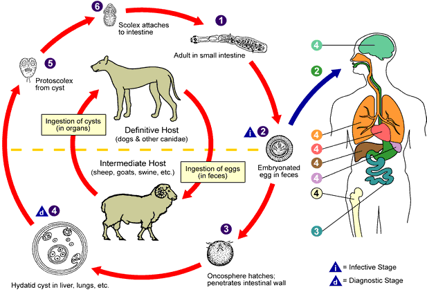
-The life cycle of E. granulosus is completed in two hosts:
- Definitive host- dogs and wild canines
- Intermediate hosts– man, sheep, cattle, goat etc.
- The eggs are discharged with the feces of the definitive hosts.
- These are swallowed by the intermediate hosts sheep and other domestic animals while grazing in the field and also by man, especially by children due to intimate handling of infected dogs.
- In the duodenum, the hexacanth embryo are hatched out.
- In about 8 hours after ingestion the embryo bore their way through the intestinal wall and are carried by the blood stream to various internal organs especially the liver and lungs are the most common sites.
- In these organs, the embryos get through the initial inflammatory response of the host and undergo dramatic changes.
- It increases in size up to 5-8 cm in few months and is finally transformed into a fluid filled hollow bladder, hydatid cyst.
- The cyst has an inner germinal layer that contains a large number of nuclei from which buds of tissue develop into hollow fluid filled brood capsules.
- These brood capsules may remain affixed to the cyst wall by means of their peduncle or may float in the hydatid fluid.
- Eventually many larvae are produced in large numbers from the germinal layer within the brood capsule.
- As the cyst grows older, a larger number of brood capsules and free protoscolices float free inside the hydatid fluid.
- Dogs and other canid host gain infection by ingestion of hydatid cyst containing protoscolices (fertile cyst), present in the viscera of sheep, cattle or other intermediate hosts.
- In the small intestine, protoscolices evaginate, penetrate deep between villi and enter the cyst of Lieberkuhn.
- It develops into a mature adult worm and began to produce infective eggs within 41-76 days of ingestion of the hydatid cysts.
- The life cycle of parasite comes to a dead end in man as dog have no access to infected viscera of man containing hydatid cyst. The natural cycle is maintained between dog and sheep.

Mode of transmission of Echinococcus granulosus:
- The disease caused by E. granulosus is zoonotic.
- Human being an accidental host, acquires the infection through ingestion of the eggs in following ways:
- Direct contact with infected dogs.
- Indirectly through food, water and other materials contaminated with eggs of the parasite.
- Less commonly, by coprophagic flies which may aid as mechanical vector of eggs.
Pathogenesis of Echinococcus granulosus:
- Mainly mechanical damage is produced.
- The young cyst that develops embryos lodged in vital centers may soon infected with functions of the organ with damaging, even fatal results.
- Benign cyst may be asymptomatic or it may produce physical burden to the patient.
- The severity relies on the type of tumor and organ or tissue where it first becomes implanted and Anaphylactic reactions develop.
Clinical manifestation of Echinococcus granulosus :
- The parasite gives rise to cystic echinococcosis earlier known as hydatid disease or hydatidosis. The hydatid cyst is primarily responsible for the pathogenesis of the disease.
- The condition persists as asymptomatic for a long period after infection in a majority of cases.
- The clinical symptoms in CE in symptomatic cases, relies upon the organ involved, interaction between the expanding cyst and adjacent organ and complications occurred by rupture of cyst.
- The cyst in vital organs interfere with the function of affected organs with the risk of fatality in certain cases.
- Hepatic hydatid:
- It is seen in 66% of cases. It may appear as hepatomegaly, with or without palpable abdominal man.
- The condition is linked with pain, nausea and vomiting, portal hypertension and biliary peritonitis.
- Cyst may rupture into bile ducts, leading to intermittent jaundice, fever and eosinophilia.
- Allergic exhibition up to anaphylactic shock may occur in case of sudden rupture.
- Pulmonary Hydatid:
- Pulmonary cyst may be present in 22% of patients.
- Cyst occurring within the lung tissue cause hemoptysis transient thoracic pain and shortness of breath.
- In case the rupture is incomplete, cyst may transform into chronic pulmonary abscess.
- The patient reports of sudden attack of cough with sputum containing frothy blood, mucus and hydatid sand.
- Brain cyst (up to 1%)
- A large cyst may lead to symptoms of increased intracranial tension (headache, vomiting and poor vision) up to epilepsy.
- Renal cyst (about 3%)
- It causes intermittent pain and hematuria.
- The hydatid sand may be present in urine.
- Hydatid cyst of the spleen, heart may present as tumor like condition or an abscess.
- Osseous cyst (about 2%)
- Osseous cyst wall has only 1 layer the germinal layer which develops first in the narrow cavity, then extend to the osseous tissue leading to:
- Erosion of large men of bone
- Destruction of bone trabecular
- Spontaneous (pathological) fractures
Complications:
- The cyst may rupture due to the trauma and during surgery into the pericardium, the bile ducts and the GI tract leads to severe clinical outcomes such as pleural effusion, pneumothorax and secondary echinococcosis of peritoneal or pleural cavity.
- A ruptured hydatid cyst leads to two risks:
- The released hydatid fluid if absorbed in the circulation bronchi, peritoneum or pleura produces a sudden anaphylactic shock which may be fatal.
- This may result in the formation of secondary echinococcosis in various parts of body due to dissemination of scolices by the circulation.
Epidemiology
- E. granulosus is cosmopolitan in distribution.
- Infection is endemic to East and South Africa, central and South America, South east and central Europe and Middle East, Russia and China.
- Annual incidence of CE per 100000 rates vary largely among these countries.
