Anatomical structure of the dicot root:
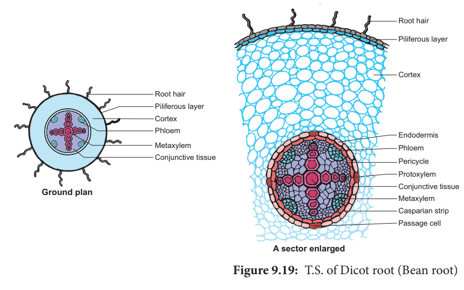
T.S. of dicot root (sunflower, Bean and pea) shows following internal structures:

Epiblema:
- It is also termed as rhizoderm or piliferous layer.
- It is outermost single layer of root which is composed of thin-walled, closely packed parenchymatous cells without intercellular spaces.
- The cuticle and stomata are absent.
- Most of epidermal cells extend out in form of tubular unicellular root hairs.
- Due to the presence of root hairs in epiblema, it is named as piliferous layer.
- This layer functions for the uptake of water and mineral salts from the soil and thus has no cuticle.
- Root hairs provide maximum surface area for absorption.
Cortex:
- It is located below the epiblema.
- It consists of many layers of thin-walled rounded or polygonal parenchymatous cells with sufficiently developed intercellular spaces between them.
- Cells of cortex consists of leucoplasts and store starch grains.
- Sometimes, outer layer of cortex becomes cutinized and forms exodermis of root.
- Cortex cells store food and conduct water from epiblema to the inner tissues.
Endodermis:
- It is the innermost layer, made up of single layer of barrel shaped compact parenchymatous cells without intercellular spaces.
- The radial walls of this layer are often thickened and sometimes this thickening extends to the inner walls also.
- Deposition of suberin and lignin causes the thickening.
- Due to deposition, strip or bands like structures are formed which are known as casparian strips or casparian bands.
- Cells of the endodermis that are located opposite the proto-xylem elements are thin-walled and termed as passage cells as they facilitate the passage of water from roots to the xylem.
- Endodermis acts as a watertight jacket around the stele.
Pericycle:
- It is located internal to the endodermis and made up of single layer of thin walled parenchymatous cells containing abundant protoplasm.
- It is very important layer as part of vascular cambium is formed from it.
- Lateral roots in dicot arise in this tissue and cork cambium also develops from it.
Conjunctive bundles:
- In between xylem and phloem bundles, there is presence of one or many layers of thin walled elongated parenchymatous cells without intercellular spaces constitutes the conjunctive tissue.
- It functions for storage of foods.
Vascular bundles:
- These are arranged in a ring but xylem and phloem form an equal number of separate bundles placed on different radii.
- As xylem and phloem are alternately arranged, the vascular bundles are termed as radial bundles.
- The number of xylem or phloem bundles varies from two to six, very rarely more.
- Xylem:
- It appears in conical in shape.
- The cells in T.S. appear polygon, and are thick walled.
- The protoxylem lies towards the periphery, so the xylem is called exarch.
- The protoxylem vessels bear annular and spiral thickenings while metaxylem vessels have reticulate and pitted thickenings.
- In mature and much developed root, the metaxylem vessels meet in centre, and pith gets obliterated.
- Xylem parenchyma and fibers are absent.
- A few tracheids are available around the vessels.
- Xylem is responsible for:
- Conduction of water and mineral salts
- Provides mechanical strength
- Phloem:
- It lies alternate to xylem patches.
- The patches are smaller and consist of sieve tubes, companion cells and phloem parenchyma.
- The phloem fibers are absent.
- The outerpart of this tissue next to pericycle is the protophloem and inner is metaphloem, but both are not easily distinguishable.
- In the hard root, a few sclerenchyma cells occur against the patch of every phloem.
Pith:
- This occupies only a small area in the center and consists of few compactly arranged, thin-walled parenchymatous cells without any intracellular space.
- Sometimes the pith is nearly obliterated owing to the wood vessels meeting in the center.
